By Eri-Tech Global Ltd · Published November 1, 2026 · Last updated November 1, 2026 · 9 min read
A modern MRI scanner room.
AI medical imaging means using software to help health staff work with pictures of the inside of the body: X-rays, CT scans, MRI scans and ultrasound images. The software does not take the picture, and it does not make the final decision. It reads an existing scan, points to patterns it has seen before, and passes that extra check to a trained person.
This article is part of the 'AI in Every Sector' series, looking at how AI is used in one sector at a time. Here the sector is health care, and the focus is narrow: what AI does around a scan, what it does well, and where a human check still matters most.
A radiographer, trained to use scanning equipment, takes it. A radiologist, a doctor trained to read images, studies it and reports for the clinician who asked for the scan. AI sits beside that pair of roles: it can sort, flag and review, but the report still goes through a person.
What AI medical imaging means
Start with the pictures. An X-ray is a quick test that uses a small dose of radiation to show dense parts of the body, such as bones, and the chest. A CT scan, short for computed tomography, uses X-rays and a computer to build cross-sectional pictures, often called slices. An MRI scan, short for magnetic resonance imaging, uses a strong magnet and radio waves, not X-rays. Ultrasound uses sound waves and a handheld device to show organs and soft parts of the body.
No single scan suits every case. A clinician asks for a scan, a radiographer takes it, and a radiologist reads it. AI enters at defined points in that chain.
AI here is machine learning: software trained on many past examples so it can spot patterns in a new image. In chest X-ray work, it has been trained to review the image and point to a possible abnormality, a part that looks different from the norm and may need a closer look. Nottingham University Hospitals NHS Trust explains in its LungIMPACT pages that AI is an extra screening tool, and it may lead to a further test, such as a CT scan, being done sooner.
The software does not look at a patient. It looks at pixels, the small dots that make up a digital image, and compares them with patterns met during training. A flag is a prompt for a person to look again, not a verdict.
A modern CT scanner, close up.
Three jobs AI does around a scan
The first job is sorting. When many scans arrive in a day, an AI tool can place the ones that look most urgent at the top of the list, so a specialist reviews them first. Bradford Teaching Hospitals NHS Foundation Trust describes this for chest X-rays: its AI support acts as a digital safety net, scanning images for subtle patterns beside its experts and flagging urgent cases for a specialist at once.
The second job is a second check. After a radiologist has read a scan, or alongside that reading, the software re-reads the same image and points to anything it thinks may have been missed. Nottingham's LungIMPACT pages call the software a second pair of eyes that re-looks at the image for extra safety. Two independent passes, one human and one software, are less likely to miss the same detail than one pass alone.
The third job is choosing the scan. Before a scan happens, someone must decide which type of scan fits the referral note, the short note that explains why the scan was requested. That step, called protocolling, is normally done by hand. In NHS Highland, a small team built a two-step AI tool for MRI referrals: first it reads the referral note and decides the general area, such as brain, spine or heart, then it narrows that down to the exact type of scan needed. Every suggestion still required a check by a scanning radiographer before a patient was scanned.
These three jobs share one pattern: the software handles a repeatable, well-defined task, and a trained person stays in charge of the step that affects the patient. When you hear a broad claim about AI in health care, ask which job, at which step, with which human check.
What is happening in the NHS
The clearest large-scale example in England is chest X-rays. On 10 June 2026, the UK government announced £20 million to roll out AI-powered chest X-ray tools to every NHS trust in England by 2029. They were already in use in about half of England's trusts, and the government said they had helped more than 4 million patients receive a faster lung cancer diagnosis or an all-clear.
Early data in the same announcement put the change in plain terms: radiologists analysed scans in an average of 4 days, compared with 8 days for the most complex cases before. The government also said the NHS carries out more than 7 million chest X-rays each year, and faster analysis should help more patients begin treatment within 62 days of a GP referral, in line with cancer waiting-time standards.
The same package added £8.1 million, through the National Institute for Health and Care Research, to test six further AI and digital technologies at 12 NHS trusts and one GP partnership across England and Scotland. Those projects cover analysis of CT scans, electrocardiograms and X-rays, digital therapy, and finding the most urgent cases first, for heart failure, stroke, lung cancer, lung infections and tic disorders.
The announcement also carries a caution from the clinical side: the president of the Royal College of Radiologists said AI should support doctors by helping them work more effectively, rather than replacing expert judgement, and that radiologists will remain central to diagnosis, clinical decision-making and patient care.
How AI learns from past scans
An AI imaging tool is only as good as the examples it learned from. Training means showing the software many past scans, each labelled by experts, until it can recognise patterns on its own. Where examples are few, results fall away, and the Highland MRI project shows both sides in one small study.
The Highland team collected 7,275 MRI referrals to train its tool. For scan types with plenty of data, early accuracy was high: 100% for heart scans, 99.9% for biliary system scans, and 98.2% for liver scans. For brain scans it was 73.6%, and for spine scans 79.7%. When two radiographers independently chose brain and spine scan types, they agreed only 70% of the time, so the software performed at a similar level to staff on those harder choices. The team named data availability as its main hurdle.
Those numbers come from one early project, not a national test. They show a useful rule: ask what a tool was trained on, how many examples covered your kind of scan, and how it was tested on unseen cases.
Training data also raises a fairness question. If past scans come mainly from one group of people, the software may fit that group best and others less well. The World Health Organization, in guidance published in January 2024, warns that health AI tools may be trained on poor-quality or biased data, and may produce false, inaccurate, biased or incomplete statements. A careful rollout checks results across different groups of patients, not just overall.
Where it still falls short
The first limit is accuracy on unusual cases. A scan that looks different from training examples, because of a rare condition or a fault in the image, is most likely to be misread. A second check helps, but it does not remove the risk. That is why Bradford states that every image is still personally reviewed and signed off by a qualified professional, and why Nottingham says patients in its study receive the usual care pathway.
The second limit is automation bias, a term the World Health Organization uses for a real habit: when software gives an answer, people may accept it without the same close look they would give their own reading. The defence is to keep the human reading independent, and to record cases where software and person disagree.
The third limit is privacy and security. The World Health Organization notes that health AI tools face cybersecurity risks that could endanger patient information, and its guidance asks governments to set standards and require independent post-release auditing when a tool is used at large scale. For a patient, the question is simple: who holds the scan data, and how long is it kept?
The fourth limit is evidence. A tool that works in a trial may not work the same way in daily use, where scanners differ and referral notes vary. Both the government announcement and the Highland project call their figures early data. Look for results from routine use, published and open to scrutiny, before treating a pilot result as settled fact.
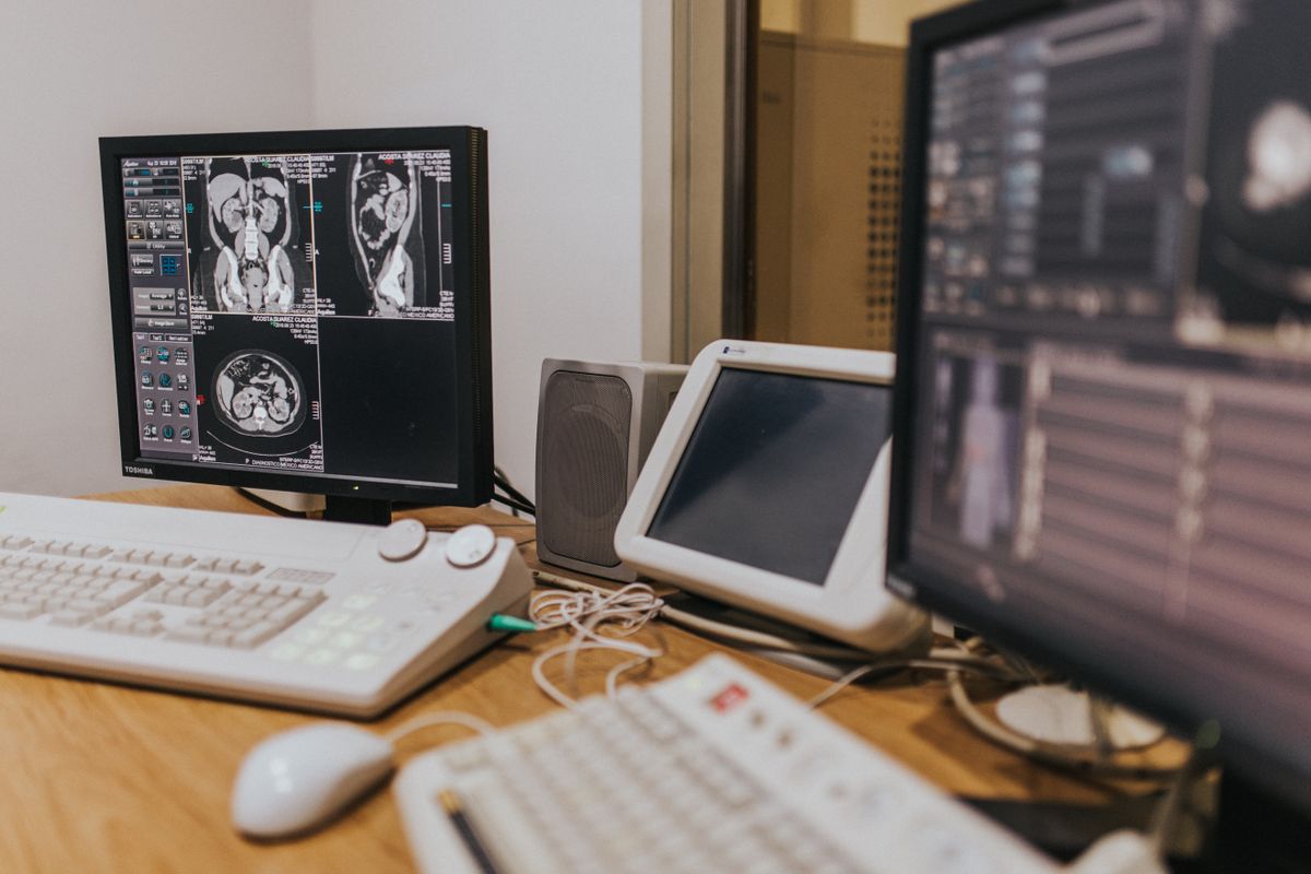
MRI scans on multiple screens.
Frequently asked questions
Does AI read my scan instead of a doctor?
No, in the examples in this article. The software reviews the image and flags patterns for a trained person, and a radiologist still reviews and signs off the result. Nottingham's study pages say patients receive the usual care pathway, and Bradford states that every image is still personally reviewed by a qualified professional.
Will AI make my scan result come back faster?
It can shorten the wait in some settings, but it does not promise a faster result for any one person. In England, early government data put average analysis for chest X-rays at 4 days, against 8 days for the most complex cases before. Your own wait still depends on the clinic, the scan type, and what further tests are needed.
Can AI get a scan wrong?
Yes. It can miss a detail, flag something that turns out to be nothing, or perform less well on scan types with few training examples. The World Health Organization warns that health AI tools may produce false, inaccurate, biased or incomplete statements, and that staff may accept a software answer too readily. That is why the human check matters more than the software on its own.
AI medical imaging is best understood as help around a scan, not as a new kind of scan. It sorts urgent cases first, gives a second check, and helps choose the right type of scan, while a trained person still reads and signs off the result. The strongest evidence so far comes from narrow, well-tested jobs like chest X-ray review.
Start with the question the tool is meant to answer, check what it was trained on, and keep a person in charge of the final report.
If you would like help turning a clear process into a simple system, explore our services or get in touch through our contact page.
This article is for general information and is not medical advice. Please speak to a qualified healthcare professional about your own situation.
What to watch next
Update this article when the NHS chest X-ray rollout reaches new trusts, when the six NIHR projects report results, or when the World Health Organization updates its AI guidance. Then update the figures and Sources together.
No. QuickBooks states that its Accounting Agent does not replace a bookkeeper or accountant, and that every suggestion must be approved before posting. The tool handles data entry and matching; your accountant handles judgement, tax rules, and advice. The ACCA survey agrees: most accountants say AI improves their work, while naming verification and human judgement as the main challenges.
Will AI bookkeeping work for a cash-heavy business?
It helps only with the records it is given. Card payments and bank transfers flow in through the bank feed, but cash sales and purchases must still be recorded by hand or photographed as receipts. A cash-heavy business should treat receipt capture as the first habit, and reconcile cash against bank deposits every month.
What should I check before trusting an AI suggestion?
Three things: the category, the amount, and the document behind it. If the category looks wrong, correct it. If the amount does not match the receipt or invoice, do not approve it. If no document sits behind the entry, find one before month end. A suggestion with no document is a guess, and guesses do not belong in a ledger.
"],["What to watch next","Update this article when Intuit or Xero move their 2026 features out of beta, change plan availability, or publish independent results for time saved. Then update the figures, the date checked, and the Sources section together, and remove or correct any claim that no longer holds."]],author:"Eri-Tech Global Ltd",metaTitle:"AI Bookkeeping for Small Firms: What It Can Do",takeaways:["AI bookkeeping handles repetitive jobs: sorting transactions, matching payments, reading receipts, and drafting invoices.","QuickBooks and Xero both added AI features in 2026 that work inside the books, with a person still approving each result.","The limits are real: AI can miscategorise a transaction, beta features vary by plan and country, and odd cases still need a human.","The safest start is small: automate one task, review its suggestions weekly, and keep your accountant in the loop."]},{title:"AI Weather Forecasting: What Is Improving",slug:"ai-weather-forecasting",category:"Technology",date:"2026-11-01",updated:"2026-11-01",primaryKeyword:"AI weather forecasting",metaDescription:"AI weather forecasting now runs beside physics-based models at ECMWF and NOAA. Learn what improved, where it falls short, and how to read a forecast well.",alt:"Dark storm clouds filling the sky, suggesting an impending storm.",image:"17845906",excerpt:"AI weather forecasting now runs beside physics-based models at ECMWF and NOAA. Learn what improved, where it falls short, and how to read a forecast well.",sections:[["What AI weather forecasting means","AI weather forecasting means using software that learns from past weather data to predict what the atmosphere will do next. It does not replace the physics-based models that forecasters have used for decades. Instead, the large weather centres now run AI models beside those physics models, compare the results, and use both when they prepare the forecast you see on your phone.","A physics-based model starts with the current state of the air, then works forward using equations for air movement, heat transfer, and rain formation. An AI model is trained on large sets of past weather records, learns how one weather state changes into the next, and applies those patterns to the latest observations. That difference explains both the speed gains and the limits below.","ECMWF, the European Centre for Medium-Range Weather Forecasts, says its AI Forecasting System, called AIFS, uses the same starting conditions as its physics-based system. Those starting conditions combine a short-term forecast with around 60 million observations from satellites, planes, boats, sea buoys, and ground stations, updated every six hours.","From that starting point, the two systems part company. The physics system, with a grid spacing of 9 km, integrates the laws of physics in its code. The AI system, with a grid spacing of 28 km in its first version, uses a model trained on past weather. A smaller grid number means smaller squares, so the model can show smaller features.","NOAA, the United States National Oceanic and Atmospheric Administration, followed a similar path. Its first AI suite grew out of Project EAGLE, a joint effort between the National Weather Service, NOAA research laboratories, the Environmental Modeling Center, and the Earth Prediction Innovation Center. The team used Google DeepMind's GraphCast model as a starting point and fine-tuned it with NOAA's Global Data Assimilation System analyses, the agency's best estimates of the current atmosphere.","That history matters. An AI model is only as good as the records it learned from, and those records come from observations and earlier physics-based work. When a rare event has few past examples, the model has less to learn from."],["What has improved so far","The first clear gain is speed. ECMWF says AIFS brings increased speed and uses approximately 1,000 times less energy for making a forecast. NOAA reports a similar pattern in its own suite. Its main AI model, the Artificial Intelligence Global Forecast System, or AIGFS, produces a single 16-day forecast using only 0.3% of the computing resources of the traditional Global Forecast System, and finishes in approximately 40 minutes.","Speed matters because a forecast is only useful if it arrives in time to act on. A faster run leaves more time to check the result, to run a second version, and to prepare a warning. It also lowers the computing bill, which is why centres can run more versions of the same forecast.","The second gain is in storm tracks. ECMWF reported on 25 February 2025, when AIFS became operational, that it outperforms state-of-the-art physics-based models for many measures, including tropical cyclone tracks, with gains of up to 20%. A tropical cyclone track is the path a storm is expected to follow. A better track helps a coastal town know sooner whether it sits in the likely path, and helps a shipping firm or an airline plan a safer route.","NOAA reported the same pattern. Its AIGFS shows a significant reduction in tropical cyclone track errors at longer lead times, so the forecast of where a storm will go stays useful further ahead. Its ensemble system, AIGEFS, which runs 31 slightly different versions to show a range of outcomes, extends useful forecast skill by 18 to 24 hours compared with the traditional ensemble system, while using only 9% of its computing resources.","The third gain is in how ensembles are combined. NOAA also runs a hybrid system, HGEFS, which joins the 31 AI members with the 31 physics members into a 62-member set. In initial testing, that combined set consistently outperformed both the AI-only and physics-only sets across most major checks. Two different methods, each with its own blind spots, can give a steadier answer together than either gives alone.","ECMWF moved in the same direction. After launching the single-forecast version, AIFS Single, in February 2025, it launched an ensemble version in July 2025. An ensemble is a group of forecasts with small differences in starting conditions. When most members agree, confidence is higher. When they spread apart, that spread is itself useful information."],["Where AI forecasts still fall short","The same reports that praise storm tracks are careful about storm strength. NOAA said its first AI version, v1.0, shows a degradation in tropical cyclone intensity forecasts, even as track forecasts improve. Intensity means how strong a storm is, including its wind speed. A forecast that places a storm correctly but understates its strength can still mislead a reader who only checks the path.","A wooden barn under dark thunderclouds.","That weakness is now being worked on in public. On 27 July 2026, the National Weather Service put an upgraded version, AIGFS v1.1, into use. Its notice says the changes improve tropical cyclone intensity forecasts, improve forecast precipitation, and reduce blurring at longer lead times. Blurring means a forecast that smooths out sharp detail as it looks further ahead, so a heavy rain band looks like a wide, light patch instead. The upgrade came from changes to the training method, including fine-tuning on four years of Global Data Assimilation System analysis.","A second limit is local detail. The World Meteorological Organization, known as WMO, noted in October 2025 that considerable challenges remain in AI systems' capability to support forecasts of local high-impact weather and hydrological processes, including floods and river flows. A global model with a 28 km grid cannot, on its own, show a single valley thunderstorm or a street-level flood. Local forecasters still need local observations, radar, and ground knowledge to turn a global forecast into a useful local warning.","A third limit is the rare event. AI models learn from the past, so an event with few past examples is harder for them to handle. That is why WMO's message is firm: AI must complement, not replace, existing forecasting methods and infrastructure. The physics models, the observation network, and the human forecaster all stay in the chain.","A fourth limit is trust in the result. An AI model can produce a confident-looking map without showing its working in the way a physics equation can be inspected. Forecasters therefore check AI output against physics output and observations. A reader can do a simpler version: compare two sources, look at the timing, and treat a single model run as one view, not a verdict."],["How to read a forecast with care","Start with the warning, not the icon. A weather app icon shows one outcome for one place at one time. A warning from a national meteorological service, or from a local authority, is the decision that matters for safety. If the app and the warning disagree, follow the warning.","Next, look at the range. Many forecast pages now show a spread of possible temperatures or rainfall amounts. A narrow spread means the models mostly agree. A wide spread means the outcome is still open, and a plan that depends on a single number is risky. For a delivery or an outdoor event, a wide spread is a signal to prepare a second option, not a reason to cancel at once.","A calm sky over the ocean at sunset.","Then check the place and the time in detail. A forecast for a city can hide big differences between a hillside and a valley, or between a coast and an inland town. Read the hourly view, not only the daily summary, when the decision involves travel, stock, or staff safety.","For a small business, turn the forecast into a short list before the season starts. Decide in advance what you will do if heavy rain is forecast for a delivery day, strong wind for an outdoor stall, or extreme heat for staff working outside. Write down who decides and how customers will be told. A better forecast helps most when the response is already agreed.","Finally, keep a simple record. Note the forecast you acted on, the warning you followed, and what actually happened. Over a season, that record shows which source you trust for your area."],["What to do next","If you run a small business, pick one weather-sensitive task this week and write its rain plan in three lines: the trigger, the action, and the person in charge. A trigger could be a heavy rain warning for your town. An action could be moving stock off the floor, confirming a delivery by phone, or shifting an outdoor job to the morning.","If you only want a better daily habit, choose one trusted source for warnings in your country and one forecast app for planning, in that order. Check the warning first, then the app for timing. When the two disagree, wait for the next update rather than picking the version you prefer.","If you work in farming, transport, energy, or construction, ask your forecast provider which models sit behind its product, and whether it shows an ensemble spread."],["Frequently asked questions","
Does AI weather forecasting replace human forecasters?
No. ECMWF and NOAA both run AI models beside physics-based models, and WMO says AI must complement, not replace, existing methods. Human forecasters check the models, add local knowledge, and issue warnings.
Why can two forecast apps disagree on the same day?
Because they may use different models, different starting data, or different ways of turning a global forecast into a local one. A disagreement is a sign of uncertainty, not proof that one app is broken. Check the warning source and the ensemble spread before you decide.
Is a faster forecast always a more accurate forecast?
No. Speed helps a forecast arrive in time and allows more versions to be run, but accuracy still depends on the observations, the training data, and the limits described above, including storm strength and local detail.
"],["Closing","AI weather forecasting has improved the speed, cost, and storm-track skill of global forecasts, and that improvement is now part of daily operations at major centres. It has not removed uncertainty, and it has not replaced the need for local warnings, careful reading, and a plan made in advance.","If you would like help turning clear, checked explanations like this into content, a website, or a workflow for your own business, explore our services or get in touch through our contact page for a free consultation."],["What to watch next","This article should be updated when NOAA issues a further AIGFS upgrade, when ECMWF reports a new AIFS version, or when WMO publishes progress on Early Warnings for All. At that point, the new figures, the date checked, and the Sources section should all be updated together, and any claim that no longer holds should be removed or corrected."]],author:"Eri-Tech Global Ltd",metaTitle:"AI Weather Forecasting: What Is Improving",takeaways:["AI weather forecasting now runs in daily operations at ECMWF and NOAA, beside physics-based models, not in place of them.","The gains so far are clearest in speed, computing cost, and the track of large storms, while storm strength remains harder to get right.","A forecast is a range of possible outcomes, not a single fixed result, so checking the timing, the place, and the warning source matters more than any single number.","For a small business, the best use of a better forecast is a simple plan made in advance: what you will move, cover, delay, or confirm when a warning appears."]},{title:"AI in Public Services: What Governments Do With It",slug:"ai-public-services-governments",category:"Politics & Policy",date:"2026-10-31",updated:"2026-10-31",primaryKeyword:"AI in public services",metaDescription:"Learn how AI is used in public services, where it helps most, what the risks are, and which checks keep a person in charge of decisions that matter today.",alt:"The National Assembly building in Belgrade with a green dome and classical columns in warm daylight.",image:"28451083",excerpt:"Learn how AI is used in public services, where it helps most, what the risks are, and which checks keep a person in charge of decisions that matter today.",sections:[["What AI in public services usually means","AI in public services means software used by a government body, or on its behalf, to help run a service that people use. That can be as small as a search tool that helps a visitor find the right form, or as large as a system that sorts a long queue of requests so staff can deal with the most time-sensitive cases first. The word AI covers several kinds of software. Some systems follow fixed rules written in advance. Others learn patterns from past data, and then use those patterns to sort, summarise, or predict. A newer kind, generative AI, can draft text or answer a question in plain words. Knowing which kind is in use matters, because each kind fails in a different way, and each kind needs a different check.","This article explains where AI in public services is most often used, what it is meant to improve, where the risks sit, and what checks a careful service puts around it. It draws on the OECD, an international body that studies how governments work, and on the UK Government's service guidance. It does not rank countries or predict what any government will do next. The point is simpler: if you use a public service, you should be able to tell when AI is involved, what it does, and how to reach a person when the answer looks wrong.","Start with the job, not the tool. A public service has a queue, a set of rules, and a record to keep. A person applies, asks, reports, or pays, and staff check the details, apply the rule, and record the result. AI enters that chain in three places: helping the person at the front find the right page or guidance, helping staff in the middle sort cases and pull records together, and helping managers at the back see where delays and errors build up.","None of those uses means the machine decides on its own. In the OECD's account, most cases used older, rules-based methods or established machine learning, which means software trained to spot patterns in data. Generative AI, including large language models, was less common. That distinction matters. A rules-based tool does what its rules say, and its mistakes can usually be traced to a rule. A pattern-based tool is harder to trace, because its answer comes from many small signals in its training data. A generative tool can write a smooth, confident answer that is still wrong, because it is built to produce likely text, not to check a fact against a record."],["Where it shows up first","The OECD report Governing with Artificial Intelligence, published on 18 September 2025, looked at 200 cases across 11 core functions of government. It found AI most common, in terms of total cases, in public service delivery and justice functions and in civic participation. Use was less common in policy evaluation, tax administration, and civil service reform. In between sat public procurement, financial management, fighting corruption and promoting public integrity, and regulatory design and delivery.","That pattern makes sense when you think about the work. Public service delivery covers many everyday tasks, so there are many routine steps that can be automated. Justice administration, the OECD notes, faces public demand and growing backlogs. Tax administration faces strict rules on how tax data may be used, which limits what a tool can be given. Policy evaluation often needs judgment about a whole programme, not a single case.","The benefits found most often were also plain. The largest share of cases aimed for automated, streamlined, and tailored processes and services. The next groups aimed for better decision-making and forecasting, and for stronger accountability and anomaly detection, which means spotting cases that do not fit the usual pattern, such as a possible error or fraud. The OECD also records an Alan Turing Institute estimate that AI could automate 84% of repetitive public service transactions in the United Kingdom, saving the equivalent of 1,200 person-years of work each year. That is an estimate of what could be automated, not what has been."],["Why the same tool needs different checks","A tool that helps a visitor find a form and a tool that helps decide whether a person qualifies for a benefit are not the same risk, even if both use similar software. The OECD puts the point plainly: there is no such thing as risk-free AI adoption. Biased algorithms, meaning software whose results treat some groups unfairly because of the data or rules behind it, can lead to adverse outcomes. Misuse of AI can infringe on, or prevent the free exercise of, human rights. Too little transparency, which means too little clear information about how a decision was reached, can weaken accountability and cause public resistance. Over-reliance, meaning trusting an output without checking it, can widen digital divides, which are gaps between people who can use digital services easily and people who cannot, and can let a single error repeat across many cases.","Those risks grow at scale. One wrong answer in a chatbot is a nuisance. The same wrong pattern applied to thousands of cases becomes a systemic error, meaning an error built into the system itself. A team that cannot explain its own tool cannot check it, or fix it quickly. There is also a risk on the other side: governments that fail to use AI miss chances to improve services. So the choice is between a tool with checks, a tool without checks, and a slow service that leaves old problems in place."],["The checks that keep a person in charge","The OECD groups what governments need into three parts: enablers, guardrails, and engagement. Enablers are the foundations, such as governance, good data, digital infrastructure, skills, and funding. Guardrails are the rules and checks that run through the life of a system, such as policies, guidance, transparency and accountability mechanisms, and oversight bodies. Engagement means ways for the public, civil society, and businesses to shape a service meant for them.","The UK Government's service guidance, updated on 15 October 2026, turns the same ideas into rules for a single service. It says a service using AI must be safe for users, protect their data, be as environmentally sustainable as possible, and stay secure. AI should only be used where there is clear evidence of a user need, and more established technology may solve the problem better. If a clearer form would fix the problem, AI is not needed.","The guidance also sets out what users must be told. If AI is used, a service must make clear how that affects a user's data and the outcome they receive. If it uses an AI-powered chatbot, users must understand that the answers do not come from a human and might not be accurate, and they must know how to contact a human being. The service must be accurate, users must be protected from bias, the service owner must understand the technology and its risks, and someone on the team must be able to explain the service's use of AI. Security professionals must be involved early, and the service must be monitored through its life, with the team prepared to stop using AI if needed. A UK Government answer given in Parliament on 11 March 2026 adds the same shape for tools handling sensitive data: the Artificial Intelligence Playbook for the UK Government and the Generative AI Framework for UK Government, both published in February 2025, stress data protection impact assessments, clear accountability, and human oversight.","Put together, those checks form a short test. Is it clear that AI is involved? Can someone explain the result? Is there a human route when the result looks wrong? Is personal data handled under a clear rule, with a check done before launch? And is the tool still watched after launch, with a way to stop it? A service that answers yes to all five is not free of risk, but it has the parts that let a mistake be found and fixed.","An office worker reviewing documents at a computer, representing everyday administrative work."],["What to watch next","The OECD is clear that many government AI efforts are still at an early stage, and that scaling up a successful pilot is hard. Skills gaps and difficulty in obtaining and sharing quality data appear across government functions. Strategies are common, but a lack of concrete guidance can stop a strategy from becoming practice. Monitoring is often too weak to show progress or spot risks, and costs are a common challenge.","The OECD's advice is to start with high-benefit, low-risk applications: cases where the gain is clear, such as cutting a backlog, and the harm if the tool errs is limited and can be caught by a person. Most governments still lack processes to measure results in full, covering spending efficiency, service quality, and potential harms. For readers, the signs to watch are simpler: does the service say, in plain words, when AI is involved? Does it keep a human route open? Is it checked over time?","A quiet public library reading room with tables and bookshelves, representing access to public information."],["Frequently asked questions","
Does AI decide whether I qualify for a public service?
It depends on the service, and the service should say. AI was most often used to automate routine steps, sort cases, and help staff find information. A careful service keeps a person responsible for a decision that affects rights or money.
What should I do if an AI answer looks wrong?
Do not rely on it alone. Use the human route the service gives, keep a copy of the answer, and check the result against the official guidance or form. The UK guidance requires an AI chatbot to tell users how to contact a human being.
Why do some public services use AI while others do not?
Because the work differs. Services with many routine cases have more places where a tool helps. Services with strict data rules, such as tax administration, or with narrow judgment work, such as policy evaluation, have fewer, according to the OECD's study of 200 cases.
"],["A careful way forward","AI in public services is best judged case by case. A tool that helps a visitor find the right form, with a person on hand, is a different matter from a tool that sorts cases affecting money or rights with no human check. The OECD's study gives a fair starting point: routine work first, clear gains, measured results, and safeguards through the life of the system. The UK guidance adds the reader's side: tell users when AI is involved, protect them from bias, keep a human route open, and be ready to stop a tool that does not hold up.","If you run a small business, use that same order online. Start with the official page, not a chatbot summary. Save the result and the date you saw it. If a step affects a payment, a licence, or a right, confirm it with a person first. For help putting a similar check into your own firm's tools, explore our services, or get in touch through our contact page."]],author:"Eri-Tech Global Ltd",metaTitle:"AI in Public Services: What Governments Do With It",takeaways:["AI in public services is most often used to automate routine steps, sort requests, and help staff find information faster.","The OECD studied 200 cases across 11 core government functions, and found use most common in public services, justice functions, and civic participation.","The main risks are biased results, unclear decisions, and too much trust in an output that no one has checked.","Good practice keeps a person in charge: users are told when AI is involved, staff can explain the result, and a human route stays open."]}, {title:"How AI Is Improving Service Pricing for Small Firms",slug:"ai-service-pricing-small-business",category:"Business",date:"2026-10-31",updated:"2026-10-31",primaryKeyword:"service pricing",metaDescription:"Learn how AI can speed up service pricing for small firms, from listing costs to break-even checks, while keeping your real costs and customers in charge.",alt:"A calculator and a blank notepad placed on paper money, with a pen beside them",image:"4386373",excerpt:"Many small firms set prices by feel. They copy a rival, round down to win a job, or add a little to last year's price and hope the maths still works. This guide explains service pricing in plain steps, and shows where an AI tool can speed up the arithmetic without taking over the decisions. At the time of writing, the pricing methods below come from guides published by the U.S. Small Business Administration and SCORE, and the AI advice stays inside those methods.",sections:[["Service pricing starts with your real costs", "Many small firms set prices by feel. They copy a rival, round down to win a job, or add a little to last year's price and hope the maths still works. This guide explains service pricing in plain steps, and shows where an AI tool can speed up the arithmetic without taking over the decisions. At the time of writing, the pricing methods below come from guides published by the U.S. Small Business Administration and SCORE, and the AI advice stays inside those methods.", "**Key takeaways**", "- Start service pricing with your own fixed and variable costs, not with a rival's price.", "- Use AI to list costs, test different prices, and compare pricing models quickly, then check every number yourself.", "- Cost-plus pricing, customer-based pricing, and competition-based pricing answer different questions, so a firm may need parts of all three.", "- A break-even check shows how many jobs you must sell at a given price before costs are covered.", "- Never treat a competitor price from an AI tool as checked. Confirm it against a real source first.", "Service pricing means choosing the selling price for a service. That choice only makes sense after you know what the service costs you to deliver. The U.S. Small Business Administration worksheet on pricing asks owners to list fixed and variable costs before they set a price.", "Fixed costs stay the same in a given period even if you sell a little more or a little less. Rent, insurance, software subscriptions, and a monthly loan payment are examples. Variable costs change with each job. Travel, materials, and pay for extra help on a busy week are examples. If you mix these two groups together, you will struggle to see why a busy month still leaves little money behind.", "AI can help here by turning a messy list into a clean table. Give it your rent, subscriptions, travel, materials, and time, and ask it to sort them into fixed and variable costs and add the totals. That work is fast, but it is not a substitute for your records. If you leave out your own pay, tax, or a regular subscription, the table will look neat and still be wrong.", "Do this first, before you ask any tool for a price. Open your bank statements, invoices, and receipts for a recent month. Write down each cost, the amount, and whether it changes with each job. Keep one line for your own pay, even if you often skip it. Also count time that does not earn a fee, such as quoting, travel, and chasing payment.", "Notes, a calculator, and a record of costs on a desk."], ["How AI helps with the arithmetic, and where it stops", "AI tools are good at repeating a calculation with different numbers. That is exactly what service pricing needs, once the costs are known. You can change the price, the number of jobs, or a cost, and see the result at once. You can also ask the tool to compare pricing models side by side, using the same cost list.", "
A short, safe prompt pattern
Give the tool only facts you have checked. A useful pattern is: here are my fixed costs for the month, here are my variable costs per job, here is the price I am testing, and here is the question I want answered. Ask it to show its working and to mark any figure it had to guess. Do not paste customer names, addresses, bank details, passwords, or contract terms into a general AI tool. Remove private details first, and keep the original records in your own files.
Ask for a table, not just a final number. A table lets you spot a cost that was dropped, a total added twice, or a price used in the wrong column. When the tool returns a result, redo one line by hand or with a calculator. If it does not match, fix the input and run it again. The goal is a price you can explain to a customer in one or two sentences.
", "
Three checks before you trust a number
First, check any outside figure. If an AI tool tells you what a rival charges, treat that as unverified until you see it on the rival's own website, price list, or shop front, at the time of writing. Tools can mix up an old price, a different service, or a different town.
Second, check that the tool used your costs, not averages from elsewhere. An average from another country or trade may have little to do with your rent, travel, or pay.
Third, check the result against common sense. If a small price change suddenly turns a loss into a large profit, look for a missing cost or a wrong unit, such as hours counted as days.
"], ["Three pricing models, compared", "The Small Business Administration describes three main pricing models, and SCORE describes similar methods. Most small firms will use one model as a base and borrow ideas from the others.", "Cost-plus pricing starts with cost and adds a markup. Markup means an amount added to cost to leave room for profit. The Small Business Administration worksheet shows how to add variable cost per unit and fixed cost per unit to reach total cost per unit, then add a markup to reach a final price. If total cost per unit is higher than the price you charge, the firm is losing money on that unit.", "Cost-plus pricing is clear and easy to check. Its weakness is that it does not ask what customers value, or what rivals charge.", "Customer-based pricing, also called value-based pricing, starts with what customers can and will pay. It asks what result the customer gets, how urgent the problem is, and what cheaper options exist. The Small Business Administration worksheet suggests research for this model, such as studying how customers make decisions and how price changes affect sales. This model can support a higher price when the service saves a customer a large amount of time or money. Its weakness is that customer views are hard to measure well, and a price based only on value can still fall below cost if costs were never listed.", "Competition-based pricing starts with rival prices. A firm looks at what others charge for a similar service and sets its price near, below, or above that level. SCORE notes that competitive pricing only looks at what rivals charge and what customers will pay, and may miss your own costs.", "A rival may have lower rent, more staff, or a different service included in the price. If you copy a rival price without checking your costs, you may win jobs and lose money on each one. Competition-based pricing is most useful as a check, after you have a cost-based price and a customer-based view.", "A practical mix often works best. Use cost-plus pricing to find the lowest price that still covers costs and leaves a margin. Use customer-based pricing to test whether a higher price fits the result you deliver. Use competition-based pricing to see where your price sits in the market. If your price is far above rivals, you need a clear reason customers can see.", "Tiered pricing, described by SCORE, fits well with this mix. It offers different versions of a service at different prices, such as a basic, a standard, and a fuller version. State the full price for each tier, and what would cost extra."], ["A worked break-even example, then your next step", "A break-even point is the sales level at which income covers costs, with no profit left over and no loss. The Small Business Administration worksheet on pricing models says a break-even check is needed for any pricing method. Its formula divides monthly fixed costs by the amount left from each sale after variable costs are paid. In plain words: fixed costs per month, divided by price per unit minus variable cost per unit, gives the break-even quantity.", "Here is a worked example only, not a real business or a market price. Suppose a small service firm has monthly fixed costs of $2,000, variable costs of $20 per job, and a price of $80 per job. The amount left per job is $80 minus $20, which is $60. Divide $2,000 by $60, and the result is 33.33 jobs, so the firm would need 34 jobs that month.", "Now ask an AI tool to test changes, using the same method. If the price falls to $70, the amount left per job becomes $50, and the break-even quantity rises to 40 jobs. If variable costs rise to $30, while the price stays at $80, the amount left per job becomes $50 again, and the break-even quantity is again 40 jobs. These tests show why a small change in price or cost can change the number of jobs you need by a large amount.", "Use the same care with rival prices. The Small Business Administration worksheet gives a simple example in which a rival sells a similar item for $10, and a firm then sells at $9.50 after weighing cost and competition. That example is a worksheet illustration, not a real market price. For your own service, find the rival price from a real, current source, note the date you checked it, and note what is included.", "There are also legal and fairness limits to keep in mind. The Small Business Administration pricing worksheet lists common mistakes to avoid, including agreeing prices with rivals, dividing markets or customers with rivals, and treating customers unfairly through illegal forms of price discrimination. Do not discuss prices with rivals in order to fix them. If a pricing question touches tax, licensing, or a regulated service in your area, get advice from a qualified professional for your own situation. This article gives general education about pricing methods, and it is not financial, legal, or tax advice.", "A person writing notes while working through figures at a desk."], ["Frequently asked questions", "
Should I start with costs, customers, or rivals?
Start with costs. Costs tell you the lowest price that still covers the work and leaves a margin. Once you have that floor, use customer research and real rival prices to test whether a higher or lower price fits.
Can an AI tool set my price for me?
No, not on its own. An AI tool can sort costs, run break-even tests, and compare models quickly. It cannot know your real costs unless you give them to it, and it cannot confirm a rival price unless you check the source.
How often should I review my prices?
Review them when costs, rival prices, or what you include in a service change, and as part of a regular check of your costs. The Small Business Administration worksheet also advises owners to monitor pricing strategies and adjust them when market conditions change. Keep a dated note of each review.
"], ["Your next step", "Start with one service this week. List its costs, test two prices with a break-even check, and confirm any rival price from a real source before you use it. If you would like help turning that price into a clear service page or a simple pricing workflow, explore our services or contact us."], ["What to watch next", "This article should be updated when the Small Business Administration or SCORE changes its pricing guides, when a pricing model described here changes in common use, or when a new, well-checked source changes the break-even method. The next article in the AI in Every Sector series will look at a different sector, and the pricing methods here will stay linked from that series so readers can return to the worked example."]],author:"Eri-Tech Global Ltd",metaTitle:"Service Pricing: How AI Helps Small Firms Set Prices",takeaways:["Start service pricing with your own fixed and variable costs, not with a rival's price.", "Use AI to list costs, test different prices, and compare pricing models quickly, then check every number yourself.", "Cost-plus pricing, customer-based pricing, and competition-based pricing answer different questions, so a firm may need parts of all three.", "A break-even check shows how many jobs you must sell at a given price before costs are covered.", "Never treat a competitor price from an AI tool as checked. Confirm it against a real source first."]},{title:"AI and Passkeys: Safer Sign-Ins Without Passwords",slug:"ai-passkeys-sign-in-security",category:"Technology",date:"2026-10-31",updated:"2026-10-31",primaryKeyword:"passkeys",metaDescription:"Passkeys replace typed passwords with a sign-in tied to your device and site. Learn how they work, where they fall short, and how to set one up safely.",alt:"A hand holding a modern smartphone with a blank screen",image:"336948",excerpt:"Passkeys replace typed passwords with a sign-in tied to your device and site. Learn how they work, where they fall short, and how to set one up safely.",sections:[["What passkeys are, in plain terms","Passwords ask you to remember a secret, type it into a box, and hope no one else has seen it. Passkeys change that deal. A passkey lets you sign in by confirming on your own phone or computer, with a fingerprint, a face scan, or a PIN, instead of typing a password that can be copied, guessed, or tricked out of you.","**Key takeaways**","- A passkey is a sign-in credential tied to one site or app, not a typed secret you share with the site.","- Your device proves it holds the passkey. Your fingerprint or face data stays on the device and is not sent to the site.","- Generative AI can now build fake login pages that look very close to real ones, which makes a sign-in with nothing to type more useful.","- Passkeys do not yet work everywhere, and you still need a recovery plan if you lose a device.","A password is a shared secret. You know it, and the site checks it. That sharing is the weak point. If the secret leaks in a breach, if you reuse it on another site, or if you type it into a page that only looks like the real site, it can be used against you.","The FIDO Alliance describes a passkey as a credential based on FIDO standards, which can live on your phone or computer, or on a hardware security key. It is tied to your account on one website or app. When you sign in, you approve the sign-in in the same way you open your device: with a fingerprint, a face scan, a PIN, or a pattern.","
Two keys, not one secret
Behind the simple tap sits a pair of linked keys. The technical name for this method is public-key cryptography. Your device creates the pair when you set up the passkey. One key is public, and the site stores that one. The other key is private, and it stays with you, on your device or in your credential manager.
That split matters. The public key on its own cannot sign you in, so a stolen site database gives an attacker public keys, not the private keys needed to pass the check. A passkey is also tied to the site it was made for, so it cannot be used on a lookalike site.
FIDO draws a line between synced and device-bound passkeys. Synced passkeys move between your devices through your passkey provider, with syncing that FIDO states is end-to-end encrypted. Device-bound passkeys stay on one device, so losing that device has a sharper cost. Know which kind you have before you rely on it.
"],["How a passkey sign-in works","A passkey sign-in has three short steps. First, you go to the site or app and choose to sign in. Second, your device asks you to confirm, in the same way you open the device. Third, your device proves it holds the private key, and the site checks that proof against the public key it already holds. No typed password travels between you and the site.","For a Google Account, Google Support lists fingerprint, face scan, and phone screen lock, such as a PIN, as the ways to confirm. Google also states that passkeys cannot be shared, copied, written down, or accidentally given to someone else, which makes them harder to hand over in a phishing attack than a password.","
Signing in on a different device
You do not always sign in on the device that holds the passkey. Google describes a common case: you sign in on a computer with a passkey held on your phone, by scanning a QR code on the computer screen with your phone and confirming on the phone. Bluetooth needs to be on for this step, and on Apple devices, iCloud Keychain needs to be on for passkeys to sync.
","
What the site sees, and what it does not see
This point is often misunderstood. The site does not receive your fingerprint or your face image. FIDO states that biometric information and its processing stay on the device, and the server only receives confirmation that the on-device check passed. Google gives the same account for its service: biometric data stays on the device and is never shared with Google.
That distinction matters. A site cannot leak a fingerprint it never received, though it can still record that you signed in, and when. A passkey changes how you prove who you are, not the rest of your account activity.
","A phone in hand, with an app open on screen."],["How AI makes fake login pages harder to spot","Fake login pages are not new. What has changed is how easy they are to build and how close they look to real pages. A report in The Hacker News on 27 October 2026, written as a contributed piece by Aranza Trevino at Keeper Security, states that generative AI makes it easier to recreate familiar login screens, copy brand styling, and tailor a phishing message to a specific person or workplace. Some fake pages also copy multi-step sign-in screens to collect one-time codes, or pass your sign-in through in real time to capture a session token.","That last trick is called Adversary-in-the-Middle, or AiTM. A fake site relays your sign-in to the real service, so you may even reach the real service while the attacker keeps a session token that lets them return without another sign-in check. The Hacker News piece also warns that filters and reputation lists can miss newly registered, short-lived fake sites, and that a valid HTTPS padlock only shows the connection is encrypted, not that the site is the one you meant to visit.","
Why a typed secret fails first
A password, and even a one-time code typed by hand, can be handed to a fake page before you notice the address was wrong by one letter. A passkey breaks that chain, because there is nothing to type. Browser checks for lookalike domains can add a second layer, but they still depend on spotting a fake page.
","
AI on both sides of the same fight
It would be wrong to present AI as only a defence. The same kind of AI that helps a browser flag a suspicious page also helps an attacker write a fluent, personal message and build a convincing copy of a login screen at speed. In a 2024 survey commissioned by the FIDO Alliance, 53% of people said they had enabled passkeys on at least one account, while only 22% said they had enabled them on every account they possibly could. Adoption is growing, but uneven.
"],["Where passkeys do not yet help","Passkeys are not a fix for every account problem. The first limit is simple: not every site or app supports them. If a site offers only a password, a passkey cannot help you on that site until the site adds support. You will still need passwords, and good password habits, for those accounts.","The second limit is recovery. A passkey is only as useful as your plan for losing the device that holds it. A synced passkey should return on a new device with the same passkey provider, according to FIDO. A device-bound passkey on a single hardware key is lost with that key, unless you set up a second key or another recovery method in advance.","The third limit is account takeover that does not need a sign-in at all. A passkey protects the sign-in step. It does not stop an attacker who already holds a live session token, or fix a weak recovery email, a shared device, or a support process that can be talked into resetting an account. No single sign-in method covers every route in.","Treat strong vendor numbers about passkeys with care. Check instead: does the site support passkeys, does your device support them, and can you still get in if you lose your phone?"],["Set up passkeys without locking yourself out","Start with one low-risk account, not your main email. Pick one where you already have a recovery email and phone number set. Add the passkey, sign out, and sign back in with it. Only then add passkeys to more important accounts.","Your operating system and browser need to be up to date. For Google Accounts, supported devices at the time of writing include computers running at least Windows 10, macOS Ventura, or ChromeOS 109, and phones running at least Android 9 or iOS 16. Supported browsers include Chrome 109 or up, Safari 16 or up, Edge 109 or up, and Firefox 122 or up. Check the current Google Support page before you start, as those numbers will change.","
Only on a device you own
Google warns that you should only create passkeys on devices you personally own and use. Once you create a passkey, anyone who can open that device can use it to reach the account. Do not create one on a shared computer, a borrowed phone, or a work device others can open.
Google also warns that a newly created passkey may take 7 days to become available at sign-in. If you already hold a trusted passkey or a physical security key, Google says you may be able to shorten that wait. Do not plan a passkey switch on the same day you need urgent access to an account. Give yourself a week of overlap, with your old sign-in method still working.
","
A recovery checklist you can copy
Use this checklist before you rely on passkeys for an important account. First, note whether each passkey is synced or device-bound. Second, add a second sign-in method you control, such as a second device or a hardware key. Third, check you can still reach your recovery email and phone number. Fourth, if you lose a device, remove its passkeys from your account settings at once. Fifth, test the recovery path once, on purpose, while calm. If a service gives no clear recovery path, keep your existing sign-in method until it does.
","Hands holding a smartphone over a wooden surface."],["Frequently asked questions","
Do passkeys replace my password everywhere?
No. Passkeys replace passwords only on sites and apps that support them. For other accounts, you still need a password, and you should keep those passwords distinct.
Will a site get my fingerprint or face scan?
No. The fingerprint or face check happens on your device. FIDO states that biometric information stays on the device and is never sent to a remote server, and Google states the same for its service. The site receives proof that the on-device check passed, not the biometric data itself.
What happens if I lose my phone?
It depends on the kind of passkey. A synced passkey should return when you set up a new device with the same passkey provider, according to FIDO. A device-bound passkey on a lost hardware key will not return on its own, so you need a second key or another recovery method in advance. Remove passkeys from the lost device in your account settings at once.
"],["A safer next step","Pick one account, check that it supports passkeys, and set up a passkey on a device you own, while keeping your current sign-in method until you have tested sign-in and recovery.","If you would like help setting up a low-risk sign-in plan for your business, explore our services or contact us for a free consultation."],["What to watch next","Update this article if a major service used by small businesses adds or removes passkey support, if Google changes its 7-day wait for new passkeys, or if FIDO publishes new adoption figures. At the time of writing, the facts above were checked on 2026-10-31."]],author:"Eri-Tech Global Ltd",metaTitle:"Passkeys: Safer Sign-Ins Without Passwords",takeaways:["A passkey is a sign-in credential tied to one site or app, not a typed secret you share with the site.","Your device proves it holds the passkey. Your fingerprint or face data stays on the device and is not sent to the site.","Generative AI can now build fake login pages that look very close to real ones, which makes a sign-in with nothing to type more useful.","Passkeys do not yet work everywhere, and you still need a recovery plan if you lose a device."]},{title:"How AI Is Changing Music Discovery and Royalties",slug:"ai-music-discovery-royalties",category:"Entertainment",date:"2026-10-30",updated:"2026-10-30",primaryKeyword:"music streaming royalties",metaDescription:"Learn how AI shapes music discovery, how music streaming royalties are shared by streamshare, and what listeners and artists should check first today.",alt:"A band performing on a stage with colourful lights in front of a crowd at a night music festival.",image:"2675648",excerpt:"Learn how AI shapes music discovery, how music streaming royalties are shared by streamshare, and what listeners and artists should check first today.",sections:[["How AI finds music you might like","You ask for music for a morning run, a quiet evening, or a long drive, and a playlist appears in seconds. That ease is one side of a larger change in music streaming royalties, because the tools that help you find a song also shape which songs get heard, and hearing is the first step toward payment.","Music streaming royalties are payments linked to the use of recorded music and songs on streaming services. This article explains how AI helps listeners find music, how the payment pool is divided, why the same number of plays can lead to different amounts, and where AI-made artist identities fit in.","AI, in this setting, means software that learns from patterns in data. A streaming service can look at songs you played fully, songs you skipped, songs you saved, and the time of day you listened. It can also look at features of the songs themselves, such as tempo, mood, and style, and at what listeners with similar tastes chose next. That mix lets a service build a playlist from a plain-language request, such as calm music for reading or upbeat music for exercise.","This is useful, and it has a clear limit. A recommendation is a guess based on past data. It can narrow your taste if you always accept the first suggestion, and it can miss music that does not fit a neat pattern, such as a new sound from a small scene. A good habit is to treat a playlist as a starting point. Save the tracks you like, follow the artists behind them, and search by artist name or album when you want to go beyond the suggestion.","For artists, discovery works in a similar way, but from the other side. A track needs clear, correct information attached to it: the artist name, the song title, the writers, and the recording owner. If that information is wrong or missing, listeners may struggle to find the track again. Good metadata, a plain word for the data that describes a recording, is therefore part of the work, not an afterthought.","A person wearing headphones and browsing a music playlist on a phone."],["Music streaming royalties: how streamshare works","A royalty pool is the pot of money in a market, in a month, that is set aside for rightsholders. A rightsholder is a person or company that owns rights in a recording or a song. Spotify explains its method, called streamshare, in simple arithmetic. Take the number of times a rightsholder’s music was played in one country in one month. Divide that number by the total number of plays in that country in that month. The result is that rightsholder’s share of that country’s royalty pool for that month.","Spotify gives a clear example. If your music is 0.1% of all streams in Mexico in a month, you get 0.1% of Mexico’s royalty pool for that month. The pool itself is based on subscription and advertising money raised in that market.","Spotify also puts the scale in context, at the time of writing. In 2025, artists who accounted for 1 in every 1 million streams on Spotify generated over $11,000, on average. That figure was about $1,000 in 2014. The same page notes that more than 400,000 songs were streamed more than a million times in 2025 alone, and that hearing all of them, one after another, would take more than 2.5 years of nonstop listening. Those figures show why a single play has no fixed price: the pool is divided by share, measured against that month’s total in that market."],["Why the same number of streams can lead to different pay","Two artists can each reach one million streams and still receive different amounts, and that is not a mistake in the maths. It follows from three steps that sit between a play and a payment. First, the market matters. A stream in a market with strong subscription income sits in a larger pool than a stream in a market where income is lower. The month matters too, because the pool and the total number of plays change from month to month.","Second, the money does not go straight from the service to the artist. Spotify pays rightsholders, and those rightsholders then pay others under their own agreements. A signed artist may be paid by a label. An independent artist may be paid by a distributor, which is a company that delivers music to streaming services and passes on the recording share, less its fee or percentage. Songwriters and publishers are paid through a separate route, which can involve collecting organisations, which are bodies that collect and distribute money for the use of songs.","Third, the contract matters. A label agreement may include an advance, which is money paid up front and later recovered from royalties, and a royalty rate that applies after that recovery. A distributor agreement may pass on a larger share, but the artist still pays the distributor’s fee. A songwriter’s share depends on who wrote the song, who owns the publishing rights, and how those rights were split. None of these details are visible from a stream count alone.","The wider market shows how large streaming has become, at the time of writing. The International Federation of the Phonographic Industry, known as IFPI, reported through Reuters that global recorded music revenues grew by 6.4% to $31.7 billion in 2025, the eleventh straight year of growth. Streaming accounted for around 70% of global recorded music income, with total streaming revenues passing $22 billion. Paid subscription streaming rose by 8.8% and accounted for 52.4% of total revenues, with 837 million paid streaming subscription accounts in total. Growth was fastest in Latin America, up 17.1%, followed by the Middle East and North Africa and by Sub-Saharan Africa, both up 15.2%, while the United States and Canada rose by 3.5%.","One limit in the IFPI report deserves a clear note. The report did not say what share of revenues record labels passed on to the artists. That gap matters for readers who want to judge fairness. A large total for the industry does not, by itself, show how much reached any single artist, because the split after the pool depends on the agreements described above. For an artist, the practical step is to read the agreement before release, and to check the first royalty statement against it, with help from a trusted adviser if the wording is unclear."],["AI-generated identities and spam tracks","AI can also create music and, in some cases, a whole performer identity. That is where listeners need a second kind of check, beyond the playlist itself. Trusted Reviews reported that Spotify is introducing an “AI persona” label for AI-generated artist identities, and that such profiles will be kept out of personalised recommendations by default. The labels were reported to appear on artist profiles and track listings from September. The company also said it would allow creators to disclose when an artist identity was created with AI, and that it would use human reviewers and AI-powered tools to spot profiles that try to hide their artificial origins.","The distinction in that policy is worth noting. It applies to AI-generated identities, rather than to AI-generated music as a whole. A human musician who uses AI tools as part of the creative process is not the same as a fully invented performer presented as if it were a real person.","Spam tracks are a related problem. Trusted Reviews reported that Spotify said it removed 75 million spam tracks from its platform over a 12-month period last year, from a catalogue of around 100 million tracks. Spam, in this context, means tracks that are low in quality, misleading, or designed mainly to game the system, rather than to serve a listener. A track that is removed, or kept out of personalised lists, cannot collect the same share of plays. IFPI chief executive Victoria Oakley called streaming fraud “theft, plain and simple”, as reported by Reuters. Fraud, in this context, includes fake plays designed to take a share of the pool without a real listener. Buying fake plays may look like a shortcut, but it risks removal and loss of trust, and it takes money from the same pool that real listeners and artists rely on.","For a listener, the check is simple. Look at the artist profile before you follow it. If a label shows that the identity is AI-generated, decide with that fact in view. If a profile has little history and many similar tracks released at speed, treat it with care."],["What to do next, whether you listen or make music","If you listen, start with one small change this week. Pick an artist you already enjoy, save three of their songs, and add them to a playlist you control. That action helps the artist’s share, and it also teaches the system about your taste in a way you can see and change.","If you make music, start with the basics before you chase a playlist place. Confirm that every track has correct artist, title, writer, and owner information. Read your distributor or label agreement, and record your songwriting splits in writing. In both cases, keep the source of truth in view. A playlist suggestion is a useful guide, not a verdict on quality. A stream count is a measure of share, not a pay slip.","A studio microphone with a pop filter in a recording studio."],["Frequently asked questions","
Does AI set how much an artist is paid per stream?
No. AI helps with discovery, but payment on Spotify is based on streamshare: the artist's share of streams in a market in a month, applied to that market's royalty pool. Contracts then decide how that share is divided.
Why do two songs with similar stream counts earn different amounts?
Because the market, the month, and the contract differ. A stream in a larger market sits in a larger pool, and a label, distributor, publisher, or collecting organisation may take a different share before the artist or songwriter is paid.
How can I tell if an artist profile is AI-generated?
Look for an “AI persona” label on the artist profile and track listings, where Spotify has applied it, and check the profile history. A human artist who uses AI tools is not the same as a fully invented performer.
"],["Closing","If you remember one point, let it be this: AI has made finding music faster and more personal, but it has not replaced the need to check how payment works, who owns the rights, and whether the performer behind a profile is real.","If you would like help turning that kind of clear, checked explanation into content, a website, or a workflow for your own business, explore our services or get in touch through our contact page for a free consultation."],["What to watch next","This article should be updated when Spotify changes its streamshare method, its AI persona policy, or its royalty reporting, or when IFPI publishes a new Global Music Report with figures that change the totals used here. At that point, the new figures, the date checked, and the Sources section should all be updated together, and any claim that no longer holds should be removed or corrected."]],author:"Eri-Tech Global Ltd",metaTitle:"Music Streaming Royalties: How AI Changes Discovery",takeaways:["AI helps discovery by matching your past listening and plain-language requests to new tracks, but it does not set the payment rate for a stream.","On Spotify, payment is based on streamshare: your share of streams in a market, in a month, sets your share of that market's royalty pool.","Contracts decide much of what finally reaches an artist or songwriter, after labels, distributors, publishers, and collecting organisations take their shares.","AI-generated artist identities are now labelled in some cases, and such profiles are kept out of personalised recommendations by default."]},{title:
Learn how AI scans food labels, what the FDA and UK formats mean, and why the printed label stays the check to use first when an app estimate looks wrong.
No. QuickBooks states that its Accounting Agent does not replace a bookkeeper or accountant, and that every suggestion must be approved before posting. The tool handles data entry and matching; your accountant handles judgement, tax rules, and advice. The ACCA survey agrees: most accountants say AI improves their work, while naming verification and human judgement as the main challenges.
Will AI bookkeeping work for a cash-heavy business?
It helps only with the records it is given. Card payments and bank transfers flow in through the bank feed, but cash sales and purchases must still be recorded by hand or photographed as receipts. A cash-heavy business should treat receipt capture as the first habit, and reconcile cash against bank deposits every month.
What should I check before trusting an AI suggestion?
Three things: the category, the amount, and the document behind it. If the category looks wrong, correct it. If the amount does not match the receipt or invoice, do not approve it. If no document sits behind the entry, find one before month end. A suggestion with no document is a guess, and guesses do not belong in a ledger.
"],["What to watch next","Update this article when Intuit or Xero move their 2026 features out of beta, change plan availability, or publish independent results for time saved. Then update the figures, the date checked, and the Sources section together, and remove or correct any claim that no longer holds."]],author:"Eri-Tech Global Ltd",metaTitle:"AI Bookkeeping for Small Firms: What It Can Do",takeaways:["AI bookkeeping handles repetitive jobs: sorting transactions, matching payments, reading receipts, and drafting invoices.","QuickBooks and Xero both added AI features in 2026 that work inside the books, with a person still approving each result.","The limits are real: AI can miscategorise a transaction, beta features vary by plan and country, and odd cases still need a human.","The safest start is small: automate one task, review its suggestions weekly, and keep your accountant in the loop."]},{title:
AI weather forecasting now runs beside physics-based models at ECMWF and NOAA. Learn what improved, where it falls short, and how to read a forecast well.
AI bookkeeping can sort transactions, read receipts and draft invoices, but it still needs a human check. Learn what it does well and where it falls short.